Histotripsy: A Revolutionary Non-Invasive Cancer Treatment
Explore the world of Histotripsy, a revolutionary non-invasive cancer treatment that uses ultrasound waves to precisely destroy target tissue. Learn about the science behind Histotripsy, its potential benefits, and the latest research findings in our comprehensive blog post.
1) Introduction to
Histotripsy
a) Welcome to the World of Histotripsy
Hello and welcome to our blog, where we will
explore into the intriguing realm of histotripsy, a new medical procedure that
is changing the way we treat numerous health disorders. We are delighted to
have you join us on this voyage of discovery and learning, whether you are a
medical professional, a researcher, or a curious reader.
Histotripsy is a non-invasive treatment that
employs concentrated ultrasonic pulses to mechanically damage specific tissue
with millimeter accuracy. This novel approach has made headlines in the medical
world, notably in the treatment of liver cancers. The FDA has awarded
HistoSonics' Edison system, the first and only histotripsy platform available
in the United States, de novo approval. This was a huge breakthrough in the
field of histotripsy, opening up new avenues for patient care.
Histotripsy, however, is not limited to liver
malignancies. This technology's potential uses are extensive and varied, with
research into its usage in treating various ailments such as kidney cancers and
even neurological problems currently underway. The possibilities are exciting,
and we'll keep you informed of any further advancements.
We hope to give you with thorough, up-to-date
information about histotripsy through our blog. We'll look at the science
underlying it, as well as the most recent study discoveries, and talk about its
possible uses and consequences for the future of healthcare. We'll also offer
field tales, including insights from prominent researchers and physicians, as
well as patient anecdotes.
We encourage you to join us as we investigate
this intriguing new area of medical technology. We hope you find our blog
educational and fascinating, whether you're here to learn, stay current, or
simply to satisfy your curiosity. So, take a cup of coffee and join me as we
explore the realm of histotripsy.
b) Definition and Overview of Histotripsy
Histotripsy is a non-thermal, non-ionizing
ablation method guided by real-time imaging. It employs concentrated ultrasound
to mechanically destroy tissue at the cellular level, resulting in acellular
debris. The name 'histotripsy' was coined in 2004 at the University of
Michigan, with 'Histo' referring to ‘soft tissue' in Greek and 'tripsy' alluding
to disintegration.
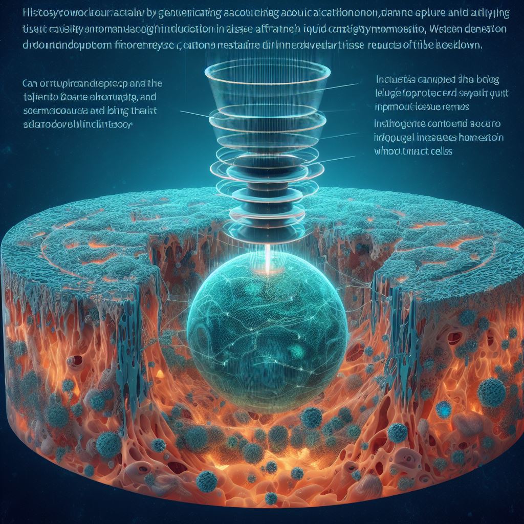
Histotripsy is based on the regulated creation
of acoustic cavitation and the interaction between cavitation and tissue, which
results in tissue destruction. When histotripsy is used on a tissue-fluid
interface (for example, blood clots or heart tissue), the tissue is eroded from
the surface inwards, resulting in holes with sharp borders. When histotripsy is
targeted inside a bulk tissue (e.g., a tumor), it liquifies the target tissue
to an acellular homogenate, and the debris is removed by the body over 1-2
months, leaving minor scars.
c) Historical Development of Histotripsy
Histotripsy was conceptualized and developed at
the University of Michigan, where most of the refining and pre-clinical
translation took place. Other organizations have lately begun to experiment
with histotripsy along both technical and translational lines of inquiry, which
serves to confirm the notion of histotripsy and enrich the potential
therapeutic utility of this novel technique.
d) Importance
and Relevance in Modern Medicine
Many pre-clinical uses of histotripsy have been
examined, including therapy for malignancies of the liver, kidney, and
prostate, neurological illnesses, thrombosis, hematoma, neonatal and fetal
congenital heart disease, valvular diseases, kidney stones, abscesses, tendons,
and biofilms. Early results from phase I human studies for histotripsy therapy
of benign prostatic hyperplasia, liver cancer, and calcified valve stenosis
demonstrate safety and viability in people.
Histotripsy is a non-invasive tumor ablation
technique that can be used to treat tumors in the pancreas, kidney, breast,
prostate, uterus, thyroid, and brain. Currently, research is concentrating on
liver and brain malignancies. In Leeds, UK, the first kidney patient was
treated with the Company's histotripsy platform, marking the first treatment in
the HistoSonics-sponsored "CAIN" Trial, which is a Phase I
prospective, multi-center study designed to evaluate the safety and technical
success of the Company's histotripsy system in targeting and destroying primary
solid renal tumors.
In animal models, histotripsy has also been
found to promote an immunological response and elicit abscopal effects, which
may have beneficial implications for future cancer treatment. Histotripsy's
non-thermal nature allows it to avoid many of the restrictions associated with
thermal devices (for example, heat sink effect, lack of accurate margins, and
predictability). Histotripsy's ability to properly remove tissue permits it to
be employed in areas where thermal methods are ineffective.
2) Mechanism of
Histotripsy
a) Fundamental
Mechanism and Process
Histotripsy is based on the regulated creation
of acoustic cavitation and the interaction between cavitation and tissue, which
results in tissue destruction. Histotripsy generates a cloud of microbubbles
(i.e., cavitation cloud) by using nano- or micrometer-sized gas pockets inside
the tissue as cavitation nuclei.
When microsecond length pulses reach negative
pressures that surpass an inherent threshold and overcome the surface tension
of existing nanoscale gas pockets, cavitation is generated during histotripsy.
Ex vivo investigations have determined that this threshold is 26-30 MPa for
water-based tissues such as blood clots, liver, kidney, heart, brain, spleen,
pancreas, and blood and water.
When histotripsy is used on a tissue-fluid
interface (for example, blood clots or heart tissue), the tissue is eroded from
the surface inwards, resulting in holes with sharp borders. When histotripsy is
targeted inside a bulk tissue (e.g., a tumor), it liquifies the target tissue
to an acellular homogenate, and the debris is removed by the body over 1-2 months,
leaving minor scars.
b) Cavitation
Cloud Histotripsy
Cavitation cloud histotripsy is the process of
initiating and maintaining a cavitation bubble cloud in order to fractionate
soft tissue. The shock scattering off one or more initial bubbles that have
developed to a suitable size in the focus is critical in establishing a thick
cavitation cloud. The shock scattering causes the positive pressure phase to be
reversed in this process, resulting in a dispersed wave with the opposite
polarity of the initial shock. The inverted shock is superimposed over the
incident negative pressure phase, resulting in an extraordinarily high negative
pressure cloud expanding toward the transducer.
Boiling histotripsy (BH) is a method that uses
High-Intensity Focused Ultrasound to mechanically fractionate solid tumors.
This approach employs a series of millisecond-long HIFU pulses comprising
shocks with high peak positive and negative pressures at the HIFU focus to
generate severe acoustic cavitation and mechanical tissue fractionation. Tissue
debris left inside a BH lesion is likely to be absorbed as part of the normal
healing process, whereas an HIFU thermal lesion will develop fibrous scar
tissue.
d) Comparison with Other Non-Invasive Therapies
Histotripsy resembles shockwave lithotripsy
(SWL) more than high intensity focused ultrasound (HIFU) in principle. The
underlying process of histotripsy, on the other hand, is fundamentally
different, depending on a mechanical action at the cellular level to damage
tissue. Histotripsy, as opposed to HIFU, is a non-thermal, noninvasive, high
precision, real-time monitoring/feedback, tissue liquefaction technology.
Many pre-clinical uses of histotripsy have been
examined, including therapy for malignancies of the liver, kidney, and
prostate, neurological illnesses, thrombosis, hematoma, neonatal and fetal
congenital heart disease, valvular diseases, kidney stones, abscesses, tendons,
and biofilms. Early results from phase I human studies for histotripsy therapy
of benign prostatic hyperplasia, liver cancer, and calcified valve stenosis
demonstrate safety and viability in people.
3) Histotripsy
Instruments and Parameters
a) Key Instrumentation Components
Histotripsy systems are made up largely of a
focused ultrasound transducer and an electronic drive system. The ultrasonic
transducer is critical for attaining the needed high focal pressure for
histotripsy. It is characterized by a large aperture, a low f-number
(transducer aperture/focal distance 1), and a high focal gain (30). The
transducer is powered by the electronic drive system, which also regulates the
ultrasonic pulses.
A complete image-guided histotripsy system also
includes an ultrasound imaging engine, an ultrasound imaging probe, a motorized
positioner or a robotic arm to move the transducer and imaging probe precisely,
and a coupling medium to ensure efficient ultrasound transmission from the
transducer to the skin. To photograph the plane containing the focal ablation
zone, the ultrasound imaging probe is normally put in the middle of the
histotripsy transducer. The system may also be guided by MRI, however an
MR-compatible ultrasonic transducer and positioner are required.
b) Parameters Used in Boiling Histotripsy
Boiling histotripsy (BH) is a kind of
histotripsy that employs millisecond-long pulses with a lower PRF. It produces
a millimeter-sized boiling bubble, which causes tissue emulsification to occur
almost instantly. At the focus, the high-amplitude acoustic wave generates a
boiling bubble, resulting in shock fronts composed of numerous high order
harmonics of the fundamental frequency. This increases energy absorption and
accelerates heating to around 100°C in milliseconds, resulting in rapid tissue
death.
Boiling histotripsy settings differ from those
utilized in other types of histotripsy. For example, the frequency employed in
boiling histotripsy is 1 to 3 MHz, the pulse length is 100 to 200 microseconds
(1-20 ms), and the pressure is 10 to 20 MPa.
c) Parameters
Used in Cavitation Cloud Histotripsy
Cavitation cloud histotripsy fractionates
tissue by creating a dense bubble cloud with microsecond-long pulses at a high
pulse repetition frequency (PRF). The parameters employed in cavitation cloud
histotripsy differ from those employed in other types of histotripsy.
Cavitation cloud histotripsy uses frequencies ranging from 250 kHz to 3 MHz,
with pulse durations ranging from 1 to 2 cycles (0.5-4 microseconds). The
needed pressure is larger than 26 MPa.
d) Safety Measures and Precautions
Histotripsy is a noninvasive focused ultrasound
method that destroys tissue by causing a mechanical action at the cellular
level. It is vital to note that the focus zone size of the ultrasonic beam,
which is defined by the frequency and geometry of the transducer, determines
the therapeutic precision of histotripsy. Multiple focal volumes are layered
together to generate the required shape and size to treat a target volume by
physically or electrically changing the focus over the ablation zone.
Cavitation can be seen on B-mode
ultrasonography as a temporally changing (twinkling), hyperechoic (bright)
zone, which is often utilized to guide histotripsy during therapy. The
histotripsy focal point is identified on the ultrasound picture and aligned
with the target tissue by moving the transducer with a robotic arm or motorized
positioner for pretreatment targeting. The targeted depth may be adjusted by
altering the transducer's depth in the coupling medium between the transducer
and the skin.
It is also critical to examine the mechanical
qualities of the tissue being treated, since these might influence the pressure
threshold required to start the cavitation bubble cloud. Increased tissue
stiffness, for example, impedes the development of early bubbles, lowering the
dispersed tensile pressure and necessitating greater beginning intensities for
cloud formation. Understanding the mechanical characteristics of the tissue can
thus aid in customizing the acoustic settings for specific tissue
fractionation.
4) Applications
of Histotripsy
Histotripsy is a non-thermal, non-ionizing
ablation method guided by real-time imaging. It employs targeted ultrasound to
mechanically destroy tissue by cavitation, converting the target into acellular
debris that is removed by the body within 1-2 months, leaving just a minor
scar.
In preclinical research,
histotripsy has been examined for a wide range of uses. These include the
treatment of malignancies in the liver, kidney, and prostate, among others. It
has also been examined for neurological illnesses, thrombosis, hematoma,
neonatal and fetal congenital heart disease, valve diseases, kidney stones,
abscesses, tendons, and biofilms.
Histotripsy was utilized to treat
primary Osteosarcoma (OS) tumors in excised canine OS samples in a feasibility
study. The findings indicated the viability of using histotripsy to treat OS
tumors, with successful cell ablation in treated regions for OS tumors and no
indication of cell death or tissue damage in normal tissues.
b) Clinical Trials and Applications
In three human clinical studies,
histotripsy was used to treat benign prostatic hyperplasia, liver cancer, and
calcified valve stenosis. Early studies indicate that people are safe and
feasible.
The HOPE4LIVER study, for
example, is a single-arm, non-randomized prospective experiment that intends to
treat all registered participants with the histotripsy device. The HistoSonics
System is designed to destroy liver tissue by histotripsy, a non-thermal,
mechanical procedure utilizing concentrated ultrasound.
c) Specific Case Studies (e.g., Osteosarcoma)
Histotripsy was utilized to treat
primary Osteosarcoma (OS) tumors in excised canine OS samples in a feasibility
study. The findings indicated the viability of using histotripsy to treat OS
tumors, with successful cell ablation in treated regions for OS tumors and no
indication of cell death or tissue damage in normal tissues.
d) Comparison with Other Non-Invasive Therapies
Histotripsy offers various
advantages over other non-invasive procedures. It is the first tumor ablation
procedure that is fully non-invasive, non-thermal, and non-ionizing. It is
capable of producing consistent and quick ablations, even close to crucial
structures. Other advantages include real-time visual guiding, excellent
accuracy, and the capacity to treat tumors of any size and form.
Histotripsy also solves the
drawbacks of thermal ablation. When administered through abdominal or
transcostal acoustic windows, it has been proven to cause constant and complete
ablation in the liver, which is extremely vascular. Histotripsy has also
demonstrated the capacity to ablate tissue around essential systems including
major arteries, bile ducts, and nerves while leaving these structures intact.
In conclusion, histotripsy is a
potential non-invasive treatment with numerous applications in both preclinical
and clinical trials. It has showed promise in the treatment of numerous cancers
and disorders, and its benefits over other non-invasive therapies make it a
viable tool for future medical treatments.
5) Advantages and Limitations of Histotripsy
Histotripsy, a non-invasive,
non-ionizing, and non-thermal ablation technology, offers several advantages:
i) Non-invasive and Non-thermal Ablation
Histotripsy employs targeted
ultrasound given from outside the body to mechanically destroy tissue by
cavitation, resulting in acellular debris. This is a non-thermal ablation
technique since it does not use heat. Because histotripsy is non-thermal, it
can overcome many of the constraints associated with thermal devices, such as
the heat sink effect, a lack of exact margins, and predictability.
ii) Real-time Imaging Guidance
Real-time imaging guides
histotripsy, allowing for accurate targeting and monitoring of the treatment
process. This feature improves the procedure's safety and efficacy.
iii) High Precision and Rapid Ablations
Even near crucial structures,
histotripsy can provide consistent and quick ablations. Because of its
excellent accuracy, it is appropriate for treating a wide range of illnesses,
including malignancies near essential organs.
iv) Ability to Treat Tumors Near Critical Structures
Histotripsy has the unusual
ability to spare tissues with enhanced mechanical strength and density,
allowing it to treat tumors near important structures while inflicting minimal
harm to these structures.
v) Minimal Remnant Scar
The debris following histotripsy
is removed by the body within 1-2 months, leaving just a minor scar. This
attribute contributes to the procedure's cosmetic and practical benefits.
vi) Stimulation of Immune Response
In animal models, histotripsy has
been found to promote an immune response and elicit abscopal effects, which may
have beneficial implications for future cancer treatment. This has the
potential to improve the body's capacity to combat cancer cells.
b) Limitations and Challenges of
Histotripsy
Despite its advantages,
histotripsy also has some limitations and challenges:
i) Tissue Properties Affecting Cavitation Threshold
Different tissues have different
resistance thresholds to histotripsy-induced damage, necessitating a different
number of ultrasonic pulses and/or pressure levels to break the tissue down.
This unpredictability can have an impact on the procedure's efficacy and
predictability.
ii) Potential Peripheral Damage
Pre-existing microbubbles, either
inside the focus region or along the intervening sound propagation channel,
might result in attenuated and dispersed treatment pulses, as well as a
distorted intended focal region. This can have a substantial impact on histotripsy's
therapeutic efficacy and may exacerbate peripheral damage outside of the
targeted target.
iii) Need for More Research
More study is needed to assess
and optimize its efficacy and safety, as well as completely investigate its
mechanism of action, pathological and immunological consequences, and the
body's short- and long-term reactions following therapy.
c) Comparison with Other Non-Invasive Therapies
Histotripsy is analogous to other
non-invasive treatments such as HIFU, radiofrequency ablation (RFA), microwave
ablation, cryoablation, and irreversible electroporation (IRE). Histotripsy, on
the other hand, is a non-thermal ablation method that depends on a mechanical
action at the cellular level to destroy tissue. This enables histotripsy to
bypass many of the restrictions of thermal devices. Histotripsy has also been
proven to trigger an immunological response, which is not typical in other
non-invasive treatments.
6) Future Directions and Research for Histotripsy
a) Optimization of Histotripsy
Histotripsy is an image-guided,
non-invasive focused ultrasound method that physically homogenizes specific
tissues. It differs from traditional thermal ablative modalities in that it is
based on the initiation and control of sonic cavitation at a target site within
the body. Histotripsy optimization is a continuing topic of study, with efforts
aimed at improving treatment efficiency and safety.
The refining of treatment systems
is a critical area of optimization. This entails fine-tuning histotripsy, which
is already recognized for its high accuracy, real-time monitoring/feedback, and
tissue liquefaction capabilities. Ultrasound backscatter analysis and shear
wave imaging, for example, have been investigated as approaches to better
determine the degree of tissue homogenization during histotripsy therapy.
The creation of ways to improve
treatment efficiency is another area of optimization. This involves using
preconditioning pulses to destroy bubble nuclei at the perimeter of the
targeted volume before delivering histotripsy pulses, which has the potential to
improve treatment accuracy.
b) Potential Future Applications and Research Directions
Histotripsy has made substantial
advances in the treatment of a variety of disorders, including liver tumors,
benign prostatic hyperplasia, and aortic valve calcification stenosis. More
study is needed, however, to completely understand its mode of action,
pathological and immunological consequences, and the body's short- and long-term
responses to therapy. According to a research that looked at the cell
composition of histotripsy sites, future uses of histotripsy might include cell
separation and transplantation. Another possible application is in cancer
treatment, where preclinical animal studies have showed promise in treating
several types of malignancies. Histotripsy is also being studied to increase
its safety, effectiveness, and adaptability. Microbubbles and fluid-filled
polymer capsules, for example, might make histotripsy safer by lowering the
energy required for cavitation. Liposomes might also be employed to deliver
therapeutic payloads, with histotripsy initiating targeted release.
c) Emerging Technologies and Innovations
Histotripsy's emerging
technologies and advancements are aimed at improving its therapeutic value and
broadening its range of uses. For example, histotripsy of the stratum corneum
may allow the diffusion of larger non-permeable biopharmaceuticals such as
heparin and insulin through skin, avoiding the need for frequent injections.
Another breakthrough is HistoSonics, Inc.'s creation of the human prototype
device (VortxRXTM) for the treatment of benign prostatic hyperplasia. In May
2013, the US Food and Drug Administration approved a human pilot trial for this
device.
Histotripsy is intended to treat
patients more safely and non-invasively in the future, with a variety of
applications that will benefit patients when additional clinical trials are
completed.
7) Ethical and Legal Considerations for Histotripsy
a) Ethical Considerations in Histotripsy
Histotripsy is a non-invasive,
non-ionizing, non-thermal ablation method that has made great success in
treating disorders such as liver tumors, benign prostatic hyperplasia, and
aortic valve calcification stenosis. When performing histotripsy investigations,
however, certain ethical factors must be taken into mind.
One of the ethical concerns is
the isolation and cultivation of high viability hepatocytes. After 1 week in
culture, histotripsy of the liver enables for the separation and culture of
hepatocytes with a high rate of viability. This is a significant topic since it
includes the manipulation of live cells, which raises ethical concerns
regarding how these cells are treated and their future use in transplantation.
Another ethical concern is histotripsy's
immunological consequences in cancer treatment. Histotripsy has been proven to
decrease the number of T regulatory cells while increasing the ratio of CD8+ to
T regulatory cells in both the tumor and the blood. This has ramifications for
the patient's immunological response and raises ethical concerns about
manipulating the immune system for therapeutic purposes.
Another ethical problem is the
use of phantoms rather than animal models throughout the early phases of
development. This is because the use of animal models in research generates
ethical concerns regarding animal welfare. However, no particular material on
the use of phantoms in histotripsy investigations was found in the search
results.
Finally, for histotripsy
investigations, ethical approval and criteria are critical. Researchers must
guarantee that their experiments are ethically sound and have acquired the
required authorisation. This includes guaranteeing the welfare of any animals
involved in the experiments, gaining informed permission from human
participants, and maintaining participant privacy and confidentiality.
b) Legal and Regulatory Aspects
In terms of legal and regulatory
issues, the FDA has awarded the Edison system and innovative histotripsy
therapeutic platforms de novo approval. Edison is the first and only
histotripsy platform accessible in the United States. The business backed up
its application for approval with data from its HOPE4LIVER studies in the
United States and Europe, which looked at the clinical safety and efficacy of
histotripsy in eradicating targeted primary and secondary liver cancers.
More study is needed, however, to
evaluate and optimize its efficacy and safety, as well as to completely
investigate its mechanism of action, pathological and immunological effects,
and the body's short- and long-term reactions following therapy. While
histotripsy has acquired regulatory approval, further study and assessment are
required to assure its safety and efficacy.
To summarize, while histotripsy
is a promising technology with tremendous promise in the treatment of a variety
of disorders, it is critical to address the ethical implications of its usage
and verify that it conforms with all necessary legal and regulatory criteria.
8) Latest FDA approval for the use of
Histotripsy for Cancer
The Food and Drug Administration
(FDA) of the United States has authorized histotripsy, a procedure that employs
sound waves to break down tumors, for the treatment of liver cancer. This
procedure was developed at the University of Michigan and represents a possible
alternative to standard cancer therapies such as surgery, radiation, and
chemotherapy, all of which can have serious adverse effects.
HistoSonics, a firm co-founded in
2009 by engineers and clinicians from the University of Michigan, received FDA
approval for the use of histotripsy to kill specific liver tissue. Since 2021,
a clinical trial at the University of Michigan Rogel Cancer Center and other
places has been treating patients with primary and metastatic liver cancers
with histotripsy. The trial proved the technology's capacity to satisfy the
major efficacy and safety goals of the testing.
HistoSonics is now able to
advertise and offer its Edison histotripsy delivery platform to hospitals and
medical practitioners for use in liver treatments. When opposed to radiation or
invasive methods, the Edison machine can ensure that the region of high
intensity ultrasound is restricted to the tumor, making it easier to ensure
that histotripsy treatments are targeting the tumor and not healthy tissue.
The histotripsy system also
includes diagnostic ultrasound imaging, which is utilized to plan and monitor
therapy in real time. Physicians may see the "bubble cloud" and how
the tissue is reacting to the treatment in real time.
Histotripsy may boost the immune
system in addition to destroying tumors. Pre-clinical research in animals imply
that the immune system learns to recognize cancer cells as dangers throughout
the cleanup process. This allows the body to continue battling the primary
tumor while also triggering a normal immune response to the malignancy.
The next step for histotripsy is
to capitalize on its immunostimulatory properties, which might be combined with
immunotherapy or medication delivery. This has the potential to transform
histotripsy from a local therapy to one that can treat tumors all over the body
and eventually lead to a cure.
9) Latest research where Histotripsy was used
a) Benign Prostatic Hyperplasia
Histotripsy has been used in
phase I human studies to treat benign prostatic hyperplasia, with preliminary
findings indicating safety and feasibility in people.
In clinical studies for the
treatment of liver cancer, histotripsy has been employed. For example, the
THERESA study, a first-in-human clinical trial, was carried out to determine
the safety and effectiveness of histotripsy in the treatment of liver cancers.
A patient with advanced and widespread metastatic disease had
histotripsy-mediated ablation of a liver lesion. There were no complications
during or after the treatment, and lab testing confirmed a considerable
reduction in the levels of the tumor marker CEA. Another clinical study for
patients with liver cancer, which took place in Barcelona, Spain, in 2018-19,
yielded promising findings.
Histotripsy has also been
utilized in phase I human studies to treat calcified valve stenosis, with
preliminary findings indicating safety and viability in people.
d) Feline Injection Site Sarcomas
A feasibility study revealed that
histotripsy ablation was feasible and well-tolerated in all three cats studied.
In all cases, precise cavitation bubble clouds were created, and hematoxylin
and eosin stained tissues demonstrated ablative damage in specified locations.
However, while histotripsy has
shown promising outcomes in the treatment of malignancies, there are still
areas that require additional investigation. Histotripsy, for example, has the
potential to liberate tumor cells from the target tumor, increasing the risk of
metastasis. As a result, further study is required to completely understand the
impact of histotripsy on the risk of recurrence and metastasis after tumor
debulking.
10) Conclusion
As we close our blog series on
histotripsy, we'd want to express our deepest appreciation to all of our
readers for their interest and participation. Your insatiable interest and
passion for information have made this voyage into histotripsy, a noninvasive,
non-ionizing, and non-thermal ablation method, a wonderfully enjoyable one.
Histotripsy, a revolutionary
technology, has showed enormous promise in the realm of medicine. It has been
studied for a variety of uses, including the treatment of cancer, neurological
illnesses, and cardiovascular diseases, with encouraging preclinical and human
clinical trial outcomes. The capacity of the device to mechanically destroy
tissue via cavitation while leaving a minimum residue scar is a significant
leap in noninvasive therapies.
Furthermore, histotripsy has been
demonstrated to activate an immunological response and create abscopal effects,
which might have a good impact on cancer treatment in the future. Recent
research has even shown that histotripsy offers the potential for effective
non-invasive tumor ablation and the prevention of local tumor development.
We are enthusiastic about
histotripsy's broad application possibilities in ablation treatment and its
ability to serve patients in the future. The current research and clinical
studies are intended to further analyze and optimize its efficacy and safety,
as well as comprehensively investigate its mode of action, pathological and
immunological impacts, and the body's short- and long-term reactions following
therapy.
We'd want to thank you for
joining us on this adventure of discovery once more. Your enthusiasm and
participation have been critical to the success of this investigation into
histotripsy. We look forward to providing you with further information and
updates on this and other fascinating medical breakthroughs. Keep an open mind,
keep educated, and, as always, thank you for reading.
FAQ’s
Histotripsy is a noninvasive,
non-ionizing, and non-thermal ablation technology guided by real-time imaging.
It uses focused ultrasound to destroy tissue at the cellular level
Histotripsy works by generating
acoustic cavitation and the interaction between cavitation and tissue, which
results in tissue breakdown. It can completely disrupt the target tissue
achieving a liquid consistency homogenate without intact cells
3)
What is the origin of the term 'histotripsy'?
The term ‘histotripsy’ was coined
at the University of Michigan in 2004. In Greek, ‘Histo’ means ‘soft tissue’,
and ‘tripsy’ refers to breakdown
4) What are the applications of
histotripsy?
Histotripsy has been investigated
for many pre-clinical applications, including treatment for tumors in the
liver, kidney, and prostate, neurological diseases, thrombosis, hematoma,
neonatal and fetal congenital heart disease, valvular diseases, kidney stones,
abscesses, tendons, and biofilms
5) What are the advantages of histotripsy
over thermal techniques?
The ability to effectively remove
tissue allows histotripsy to be used in applications that are not possible with
thermal techniques. The non-thermal nature also enables histotripsy to overcome
many of the limitations associated with thermal devices (e.g., heat sink
effect, lack of precise margins, and predictability)
6) What are the different forms of
histotripsy?
Three forms of histotripsy have
been identified, depending on the number of acoustic cycles used and the peak
negative pressures (P–) amplitudes required: intrinsic threshold histotripsy
(1–2 cycles, P– > 26 MPa), shock scattering histotripsy (3–10 cycles, P– =
15–25 MPa) and boiling histotripsy (longer 1–20 ms pulses, P– = 10–20 MPa)
7) What is the precision of histotripsy
treatment?
The treatment precision of
histotripsy is determined by the focal zone size of the ultrasound beam, which
in turn depends on the frequency and geometry of the transducer. A typical
focal zone for a spherical section aperture of 1 MHz is an elliptical shape of
1–2 mm short axis and 2–4 mm long axis
8) What are the key components of a histotripsy system?
The key instrumentation
components that make up histotripsy systems are a focused ultrasound transducer
and an associated electronic driving system
9) How is histotripsy guided during treatment?
Ultrasound imaging is typically
used to guide histotripsy during treatment as cavitation can be visualized on
the imaging
10) What happens to the tissue after
histotripsy treatment?
When histotripsy is applied to a
tissue-fluid interface, the tissue is eroded from the surface inwards creating
perforations with sharp boundaries. When targeting histotripsy inside a bulk
tissue, histotripsy liquifies the target tissue to an acellular homogenate, and
the debris is absorbed over 1–2 months by the body, leaving small scars
11) Does histotripsy affect all tissues equally?
Different tissues have specific
resistance thresholds to histotripsy-induced damage, requiring variable numbers
of ultrasound pulses and/or ultrasound pressure levels to break down the
tissue. Mechanically strong collagen-based tissues have a higher ultimate
tensile strength compared to more parenchymal structures
12) Has histotripsy been used in human
trials?
Phase I human trials have been
undertaken for histotripsy treatment of benign prostatic hyperplasia, liver
cancer, and calcified valve stenosis, with early results suggesting safety and
feasibility in humans
13) Can histotripsy increase the metastatic
rate?
There is ongoing research to
determine whether histotripsy treatments can increase the metastatic rate
14) What are the effects of histotripsy on the immune system?
Histotripsy has been shown to
stimulate cancer-specific lymphocyte responses in preclinical studies
15) Can histotripsy be used in the
treatment of brain tumors?
Histotripsy is being explored as
a potential treatment for glioblastoma and other brain tumors. It has been
shown to be tissue selective and can preserve vasculature. However, more data
needs to be obtained on the effects of histotripsy at varying ablation
parameters
16) What are the potential side effects of
histotripsy?
There can be some swelling and
bleeding following histotripsy in animal models. Further studies are needed to
evaluate its safety in humans
17) What is the future of histotripsy?
Histotripsy has broad application
prospects in ablation therapy and will benefit patients after more clinical
trials are conducted in the future
18) What are the limitations of
histotripsy?
More research is needed to fully
explore its mechanism of action, pathological and immunological effects, and
the short-term and long-term reactions of the body after treatment
19) Can histotripsy be used in combination
with other treatments?
Histotripsy can be used in
combination with other treatments such as immunotherapy. However, more research
is needed to determine the optimal parameters for immune stimulation and to
validate histotripsy for use in sensitizing resistant cancers to immunotherapy
Phase I clinical trials have
demonstrated the safety and efficacy of histotripsy in the treatment of benign
prostatic hyperplasia, liver cancer, and aortic valve calcification stenosis.
However, more research is needed to evaluate and optimize its efficacy and
safety


Comments
Post a Comment